Flow Cytometry (FCM) is a means of quantitative analysis and sorting of chemical components on the surface and inside of single-cell or other biological particle membranes. It can analyze tens of thousands of cells at high speed and simultaneously Compared with traditional fluoroscopy, it has many parameters measured in a cell, and it has the characteristics of high speed, high precision and good accuracy. It has become the most advanced cell quantitative analysis technology in the modern era. Since the 1970s, with the continuous improvement of the level of flow cytometry, its application range has become increasingly widespread. At present, flow cytometry has been widely used in clinical medicine and basic medicine fields such as immunology, hematology, cell biology, cytogenetics, biochemistry and oncology.
Flow cytometry integrates electronic technology, computer technology and fluid theory into one, which is a very advanced testing instrument. The development of flow cytometry began in 1953, when Parker and Horst designed a complete hemocytometer device that became the prototype of a flow cytometer. The principle is to first stain the whole blood cells, the white blood cells are stained blue, the red blood cells are still red, and when the cell suspension passes through the detection of the thin glass tube, it is irradiated with a beam of light, and the blue or red light emitted by different cells is filtered. After the film is converted into an electrical signal by two photoelectric converters, respectively, and then counted by the counting circuit.
Flow cytometry consists of five parts: flow chamber and liquid flow drive system, laser light source and beam shaping system, optical system, signal detection and storage, display, analysis system and cell sorting system.
The basic principle of flow cytometry is to stain the cells to be tested to make a single cell suspension. Pressing the sample to be tested into the flow chamber with a certain pressure, the cell-free phosphate buffer is sprayed from the sheath tube under high pressure, and the inlet direction of the sheath tube is at an angle to the sample flow to be tested, so that the sheath liquid can be wound The sample flows at a high speed to form a circular stream, and the cells to be tested are arranged in a single row under the coating of the sheath fluid, and sequentially pass through the detection area. Flow cytometry usually uses a laser as an excitation source. The focused and shaped beam is vertically irradiated onto the sample stream, and the fluorescently stained cells generate scattered light and excite fluorescence under the illumination of the laser beam.
These two signals are simultaneously received by the forward photodiode and the photo-multiplier tube (PMT) in the 90-degree direction. The light-scattering signal is detected at a forward angle of 10.5-2.0 degrees. This signal basically reflects the cell volume; fluorescence signal The receiving direction is perpendicular to the laser beam and is separated by a series of dichroic mirrors and bandpass filters to form a plurality of fluorescent signals of different wavelengths. The intensity of these fluorescent signals represents the intensity of the surface antigen of the cell membrane measured or the concentration of the substance in the nucleus. After being received by the photomultiplier tube, it can be converted into an electrical signal, and then converted into a continuous electrical signal by an analog-to-digital converter. A digital signal that is recognized by a computer.
The computer collects the measured signals for calculation, displays the analysis results on a computer screen, prints them, and stores them on the hard disk in the form of data files for later query or further analysis. Sorting of cells is achieved by isolating droplets containing single cells. An ultra-high frequency piezoelectric crystal is arranged on the nozzle of the flow chamber, and vibrates after charging to break the discharged liquid stream into uniform droplets, and the cells to be tested are dispersed in the droplets. The droplets are charged with different positive and negative charges. When the droplets flow through a deflection plate with several kilovolts, they deflect under the action of a high voltage electric field and fall into the respective collection containers. The uncharged droplets fall into the droplets. A waste container in the middle to achieve cell separation.
Flow cytometry plays an important role in immunology, hematology, cell proliferation and tumor pathology, apoptosis and flow karyotyping.
Dove Prism
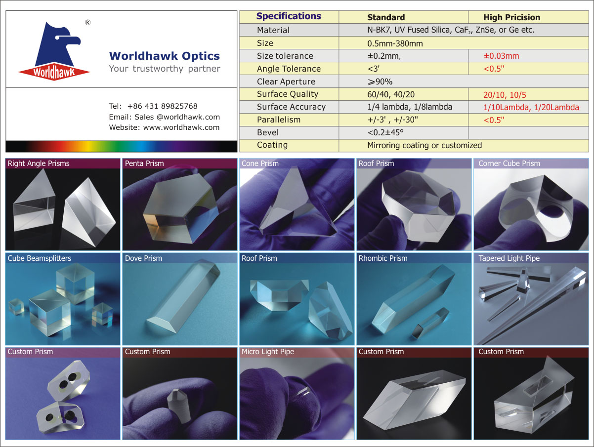
Dove prism is used to rotate, invert, or retroreflect an image, depending upon the prism's rotation angle and the surface through which the light enters the prism. Dove Prisms are fabricated from N-BK7 glass for high transmission from the visible to the near-infrared spectral range.
Dove prisms can be thought of as right-angle prisms with the triangular apex removed, which reduces the weight of the prism and stray internal reflections. They introduce astigmatism when used with converging light, so we recommend using them with collimated light.
Image Rotation
Light is usually propagated along the longitudinal axis of a Dove prism. In this geometry, light reflects once from the bottom face, inverting the image on the other side. Rotation of the prism about the longitudinal axis rotates the image at twice the rate of the prism's rotation. For example, a 20° rotation of the prism results in a 40° rotated image. The AR-coated Dove prisms are designed specifically for the image rotation and inversion application.
Due to the high incidence angle, the light reflecting from the bottom face undergoes total internal reflection, even if the light's propagation axis and the prism's longitudinal axis are not exactly parallel. Hence, in a Dove prism, the magnitude of the internal transmission is limited only by absorption.
Retroreflection
When light is incident on the longest face, the Dove prism acts as a retroreflector or a right-angle prism. The light exits parallel to the input light (independent of the incidence angle) and is inverted by 180°. In situations with limited space or where more convenient mounting options are needed, the Dove prism can replace a retroreflector or right-angle prism.
Custom Coatings
Upon request, our prisms can be AR coated for the UV spectral range , VIS spectral range and IR spectral range.

Dove Prisms,Custom Glass Prisms,Glass Trapezoidal Prism,Dove Prisms Glass Trapezoidal Prism
ChangChun Worldhawk Optics Co.,Ltd , https://www.worldhawk-optics.com
